


 Română
 Русский


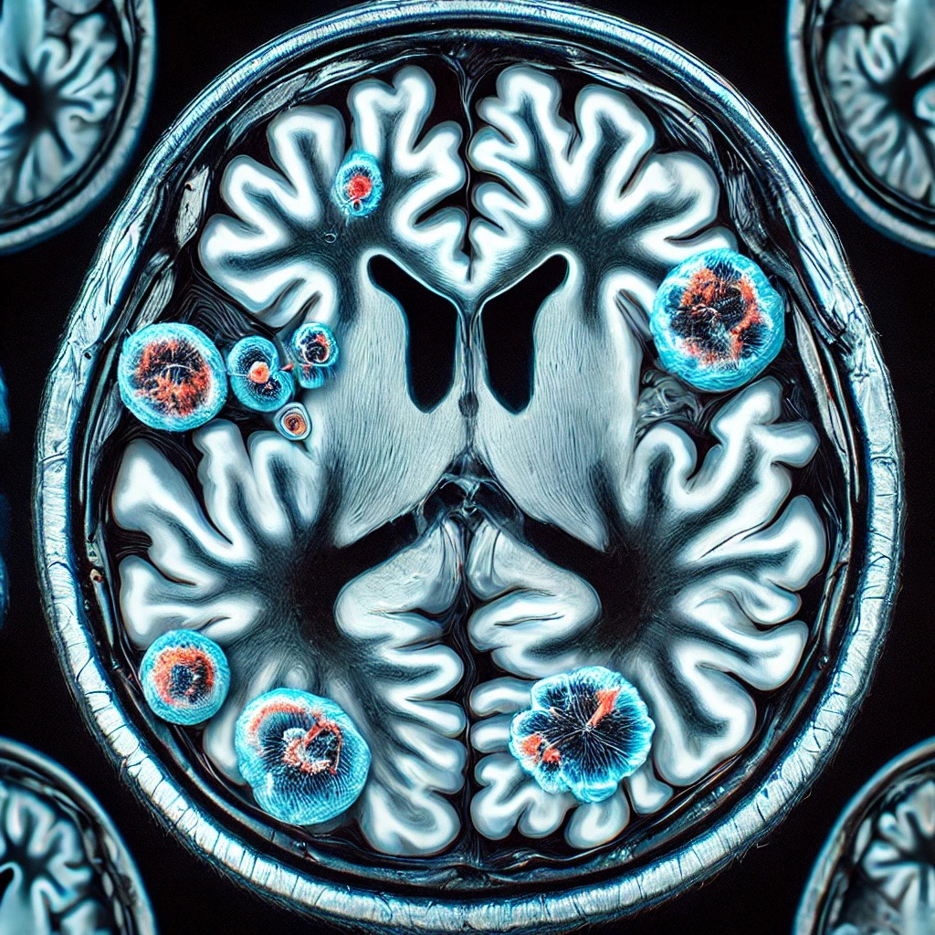
MRI 3T and Multiple Sclerosis Diagnosis: How Advanced Technology Makes a Difference
Multiple sclerosis (MS) is an autoimmune disease that affects the central nervous system, causing a variety of neurological symptoms. Accurate and early diagnosis is crucial for effectively managing this condition. At the German Diagnostic Center (CDG), we use advanced 3T MRI technology to provide detailed and clear images essential for identifying lesions specific to multiple sclerosis. In this article, we will explore how 3T MRI enhances the process of diagnosing and monitoring multiple sclerosis.
What is Multiple Sclerosis?
Multiple sclerosis is characterized by the deterioration of myelin, the protective sheath of nerve fibers in the central nervous system. This deterioration disrupts communication between the brain and the rest of the body, resulting in symptoms such as muscle weakness, coordination problems, vision disturbances, and balance difficulties.
The Role of 3T MRI in Diagnosing Multiple Sclerosis
- Early Detection of Lesions
- 3T MRI is extremely sensitive in detecting small demyelinating lesions, which are characteristic of multiple sclerosis. These lesions can be difficult to observe with older equipment, but 3T MRI provides high-resolution images that allow for their identification at early stages.
- Evaluating Disease Activity
- 3T MRI can highlight active and inactive lesions. Active lesions appear as contrasting areas after the administration of gadolinium, a contrast agent used during scanning. This ability to distinguish between active and inactive lesions is essential for evaluating disease activity and treatment efficacy.
- Monitoring Disease Progression
- Through detailed and periodic images, 3T MRI allows doctors to monitor the progression of multiple sclerosis over time. This helps in adjusting treatment according to the disease's evolution and identifying any new lesions that may appear.
How is a 3T MRI Scan Conducted for Multiple Sclerosis?
Preparing Patients: Before the scan, patients are instructed to remove any metal objects and are given details about the procedure. Examination with the administration of a contrast agent is recommended to improve visualization of active lesions.
During the Scan: Patients are positioned on a table that moves inside the 3T MRI scanner. It is important to remain still to obtain clear images. The scan can last between 15 and 30 minutes, depending on the complexity of the investigation.
After the Scan: The obtained images are analyzed by a specialized radiologist who will identify and describe the lesions characteristic of multiple sclerosis. The results are then discussed with the patient and the treating physician to decide on the next steps in managing the disease.
The 3T MRI technology available at the German Diagnostic Center represents a significant advantage in diagnosing and monitoring multiple sclerosis. Through detailed and precise images, 3T MRI provides doctors with the necessary information to give patients the best chances of effective disease management.
For more information or to schedule an investigation, please contact the Call Center operators at: 022840000.
Your health is our priority!