


 Română
 Русский


3T MRI Spectroscopy of the Cervical Spinal Cord with Contrast
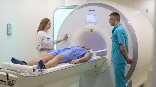
Price: 5000 MDL
3T MRI Spectroscopy of the Cervical Spinal Cord with Contrast is a unique analytical technique used to determine metabolic information for characterizing brain lesions. It allows differentiation between tumoral and non-tumoral lesions, tumor recurrences, malignant transformations, hypoxic or ischemic lesions, and helps distinguish inflammatory, infectious, and demyelinating pathologies.
In particular cases, especially when inflammation or tumors are suspected, the examination should be supplemented with intravenous administration of contrast agents. This enhances the visibility of various structures, improving image quality.
Examination
Spectroscopy is a technically complex imaging investigation that requires specialized processing and interpretation. It is primarily used for the evaluation of brain tumors, comparing the chemical composition of normal tissue with that of abnormal cells. This test is mainly used to determine the type of tumor, its aggressiveness, and to differentiate between tumor recurrence and radiation necrosis.
Preparation for the Procedure
Spectroscopy does not require special preparation or a rehabilitation period afterward, so you can continue your daily activities as usual. There are no dietary restrictions unless specified otherwise by your specialist. Women are advised to remove makeup before the examination, as some cosmetic products may contain metallic powders that can interfere with the imaging.
Before the procedure, the medical assistant will ask you to remove all metal objects (glasses, watches, magnetic cards, jewelry, etc.), as they can be quickly attracted by the magnetic field. It is mandatory to inform your doctor if you have any metallic implants (cardiac stents, pacemakers, IUDs) since these may malfunction during the MRI and pose a risk to your life.
It is helpful to bring any medical records related to previously diagnosed conditions.
Recommended Related Services
Service Name
Service Price
650 MDL


700 MDL